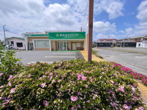
上里町の彩の丘クリニックです。 内科 消化器内科 外科 水曜と日曜休診 院内処方 神保原 駅徒歩7分 消化器病学会および消化器内視鏡学会専門医
ようこそ、彩の丘クリニックのホームページへ。
安心の医療を目指しています。技術はやさしさ、安心と安全をモットーに地域の皆様への医療貢献、そして日本の明日への医療技術の貢献を目指します。
information
気温が急上昇してきました。まだ高温に体が慣れていない時期で熱中症に注意しましょう。暑くない時間帯に 運動し、また感染症に注意しましょう。
来院の際はマスクを着用をお願いいたします。
令和5年3月13日以降のマスク着用について
厚生労働省は主に次の3カ所についてマスクの着用を推奨しています。
①医療機関受診時
②高齢者施設への訪問時
③混雑した公共交通機関利用時
7月29日(水)休診(毎週水曜休診)
8月1日(土)午後は5時まで診療
8月2日(日)休診
8月5日(水)休診(毎週水曜休診)
8月9日(日)から16日(日)まで夏季休業となります。ご不便をおかけしますがよろしくお願い申し上げます。
(毎週水曜・日曜・祝日は休診です)
リンク View and download the article
|
||||||||
|
||||||||
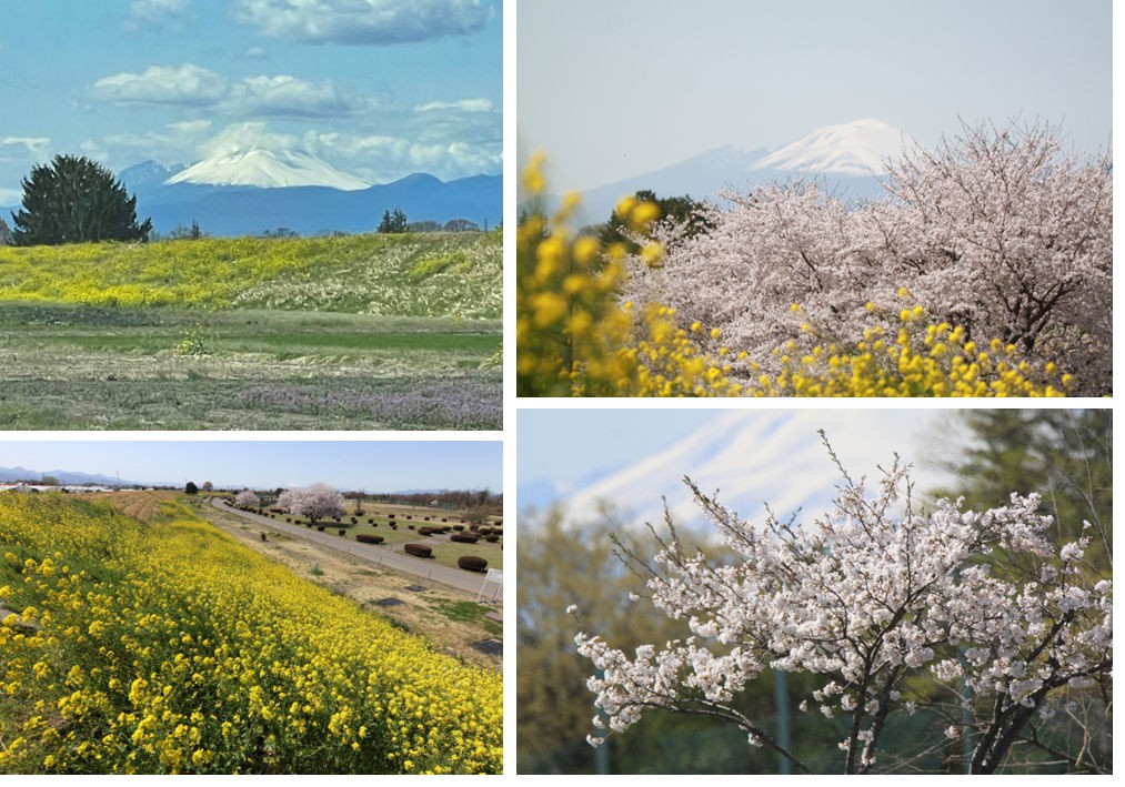
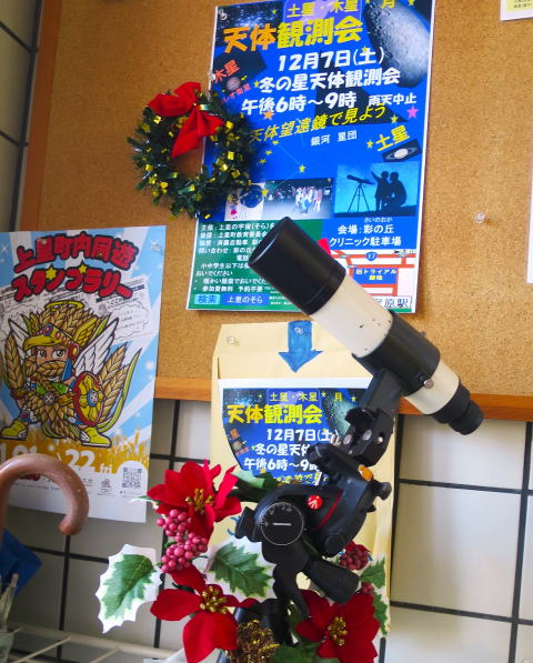
員会の後援を頂き、毎年、星空が澄み渡る10-12月に行っています。今回は11回目となりました。 詳細は 上里のそら(宇宙)を見る会ホームページをご覧ください。 |
||||||||
|
||||||||
|
||||||||
|
||||||||
|
||||||||
|
||||||||
|
||||||||
|
||||||||
|
||||||||
|
||||||||
|
||||||||
|
||||||||
|
||||||||
古い写真は下記 四季彩の丘 Web日記写真集 に移動しました。 クリックすると移動します。 |
||||||||


369-0305
埼玉県児玉郡
上里町神保原416-5
電話 0495-71-7166
■診療時間■
午前 9時-12時
午後 3時-6時
(土曜は5時まで)


*US はUltra Sound(超音波検査)の略です。


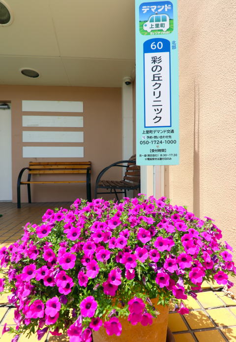
 お安く便利")


を見る会 事務局")
